
Fetal Echocardiography in Bengaluru
A detailed ultrasound of your unborn baby's heart that detects heart defects and rhythm problems before birth, so the right care is ready from day one. Under the care of Dr. Bharath A. P.
- Ideal at 18–24 Weeks
- Safe for Mother & Baby
- Detailed Prenatal Counselling
- Delivery Planning Support

Dr. Bharath A. P.
Pediatric Interventional Cardiologist
Clinical Practice
Dedicated to newborn & child cardiac excellence
Children Evaluated
Evaluated with echocardiogram, from preterm infants to young adults
Children Operated
Cardiac interventions & cath lab procedures in children
Surgeries Coordinated
Coordinated surgical care for children with congenital heart diseases.
What Is Fetal Echocardiography?
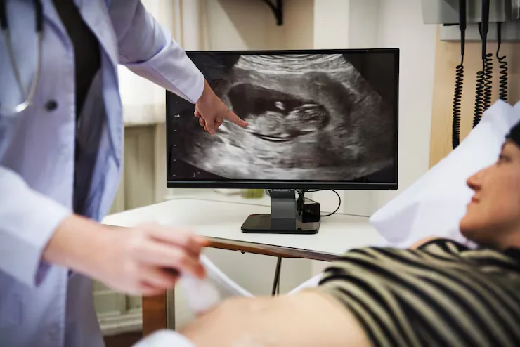
Fetal echocardiography is a specialised ultrasound scan focused entirely on the baby's developing heart. It is performed through the mother's abdomen, just like a routine pregnancy scan, but examines the heart chambers, valves, great arteries, veins and heart rhythm in much greater detail.
Detecting a heart defect before birth allows parents to understand the condition, ask questions and plan delivery at a hospital with the right pediatric cardiac support. For many conditions, this early planning makes the first days of life safer for the baby.
Structural Assessment
Four-chamber view, outflow tracts, three-vessel view and aortic arch are examined systematically.
Rhythm Evaluation
Detection of fast, slow or irregular fetal heart rhythms, including heart block.
Parent Counselling
Clear explanation of findings, likely treatment after birth and the outlook for your baby.
When Is It Recommended?
Your obstetrician may recommend a fetal echo if any of these apply:
- An unclear or abnormal heart view on the routine anomaly scan
- A previous child, parent or sibling with congenital heart disease
- Diabetes before pregnancy or poorly controlled diabetes
- Pregnancy conceived through IVF or other assisted reproduction
Not sure if your child needs this?
Share your child's reports and Dr. Bharath's team will guide you on the next step.
Conditions Covered
Heart conditions commonly assessed or treated with this service.
Septal Defects
Larger VSDs and atrioventricular septal defects (AVSD).
Outflow Tract Anomalies
Tetralogy of Fallot, transposition of the great arteries and double outlet right ventricle.
Single-Ventricle Hearts
Hypoplastic left or right heart, tricuspid atresia and related conditions.
Obstructive Lesions
Critical aortic or pulmonary stenosis and suspected coarctation of the aorta.
Rhythm Disorders
Fetal tachycardia, bradycardia and congenital heart block.
Heart Function Problems
Cardiomyopathy, fetal heart failure and fluid build-up (hydrops).
How It Is Performed
What happens on the day, explained simply so you know what to expect.
- 1Step 01 / 04
History Review
Pregnancy details, previous scan reports and family history are reviewed.
- 2Step 02 / 04
Abdominal Scan
Gel is applied to the abdomen and the baby's heart is examined from multiple angles.
- 3Step 03 / 04
Doppler & Rhythm
Blood flow through valves and vessels and the baby's heart rhythm are assessed.
- 4Step 04 / 04
Counselling
Findings are explained, and if needed, a follow-up scan and delivery plan are discussed with you and your obstetrician.
Benefits, Risks & Care
A balanced overview, plus how to prepare and what recovery looks like.
Key Benefits
- Early diagnosis allows planned, safer delivery
- Time for parents to understand and prepare
- Immediate treatment can be arranged after birth if needed
- Reassurance when the heart is found to be normal
Every child is different. Specific risks, benefits and alternatives for your child are discussed in detail during consultation.
Why Families Choose Dr. Bharath A. P.
Super-speciality training, academic experience and hands-on cath lab expertise.
National Board super-speciality fellowship at Madras Medical Mission Hospital, Chennai.
Assistant Professor of Pediatric Cardiology at Sri Jayadeva Institute of Cardiovascular Sciences and Research since 2015.
Diagnostic catheterisation and interventions including ASD, VSD and PDA closures, valvuloplasties and stenting.
Co-author of peer-reviewed articles in Heart Lung Circulation, Archives of Disease in Childhood and Annals of Pediatric Cardiology.
Frequently Asked Questions
Answers to common questions about Fetal Echocardiography.
When is the best time to do a fetal echocardiogram?
+
The ideal window is between 18 and 24 weeks of pregnancy, when the heart is large enough to see clearly. It can also be done later in pregnancy if a concern arises.
Is fetal echo safe for my baby?
+
Yes. It uses the same ultrasound technology as routine pregnancy scans, and there is no known harm to the mother or the baby.
Can a fetal echo detect every heart problem?
+
It detects most major heart defects, but very small holes and conditions that depend on changes after birth — such as a PDA or mild coarctation — may only be confirmed with an echo after delivery.
What happens if a heart defect is found?
+
Dr. Bharath will explain the condition, how it is usually treated, and the outlook. Together with your obstetrician, a plan is made for delivery at a hospital equipped to care for your baby's heart immediately after birth.
Do I need a referral for a fetal echo?
+
A referral from your obstetrician is helpful, as it explains why the scan is needed, but it is not essential. Please bring all your previous pregnancy scan reports.
Will I need more than one fetal echo?
+
If the heart looks normal, one scan is usually enough. If a problem is found, follow-up scans may be advised to monitor it and plan for delivery.
Visit the Clinic
VEGA CURRE Multispeciality Clinic
2nd Floor, Shop No. 24 & 25, 3rd A Cross, Bhuvaneshwari Nagar, Dasarahalli Main Road, Hebbal Kempapura, Bengaluru – 560024