Transthoracic Echocardiography (TTE) in Bengaluru
A painless, radiation-free heart ultrasound that shows the structure, pumping function and blood flow of your child's heart in real time. Under the care of Dr. Bharath A. P.
- Zero Radiation, No Needles
- Newborns to Adolescents
- 2D, Colour & Spectral Doppler
- Same-Visit Explanation of Results

Dr. Bharath A. P.
Pediatric Interventional Cardiologist
Clinical Practice
Dedicated to newborn & child cardiac excellence
Children Evaluated
Evaluated with echocardiogram, from preterm infants to young adults
Children Operated
Cardiac interventions & cath lab procedures in children
Surgeries Coordinated
Coordinated surgical care for children with congenital heart diseases.
What Is a Transthoracic Echocardiogram?
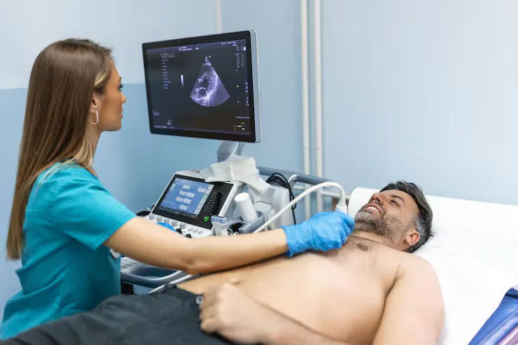
A transthoracic echocardiogram (TTE) is an ultrasound scan of the heart performed by placing a small probe with warm gel on the chest. Sound waves create moving pictures of the heart chambers, valves and major blood vessels, while Doppler imaging shows the direction and speed of blood flow.
TTE is the most important first test in pediatric cardiology. In most children it can confirm or rule out a heart defect at the first visit, measure how well the heart is pumping, and estimate pressures inside the heart and lungs — without any pain, injection or radiation.
Complete Anatomy
Systematic views of all four chambers, valves, septum, aortic arch and pulmonary arteries.
Blood-Flow Mapping
Colour and spectral Doppler detect leaks, narrowings and abnormal shunts between chambers.
Child-Friendly
Parents stay beside the child; cartoons, feeds and patience replace sedation in most cases.
When Is It Recommended?
Your pediatrician may refer your child for an echocardiogram if any of the following are noticed:
- A heart murmur heard during a routine check-up
- Bluish lips or nails (cyanosis) or low oxygen saturation
- Fast breathing, sweating or tiring during feeds, or poor weight gain
- Chest pain, fainting or palpitations during exercise
Not sure if your child needs this?
Share your child's reports and Dr. Bharath's team will guide you on the next step.
Conditions Covered
Heart conditions commonly assessed or treated with this service.
Holes in the Heart
Atrial septal defect (ASD), ventricular septal defect (VSD) and patent ductus arteriosus (PDA).
Valve Problems
Narrowing (stenosis) or leakage (regurgitation) of the pulmonary, aortic, mitral or tricuspid valves.
Complex & Cyanotic Defects
Tetralogy of Fallot, transposition of the great arteries and single-ventricle hearts.
Heart Muscle Disease
Dilated and hypertrophic cardiomyopathy, myocarditis and reduced pumping function.
Coronary & Pericardial Disease
Coronary artery changes in Kawasaki disease and fluid around the heart (pericardial effusion).
Pulmonary Hypertension
Non-invasive estimation of lung artery pressure and right heart strain.
How It Is Performed
What happens on the day, explained simply so you know what to expect.
- 1Step 01 / 04
Settling In
Your child lies on the bed or on a parent's lap. Infants can be fed and toddlers can watch cartoons.
- 2Step 02 / 04
Gel & Probe
Warm gel is applied and a small probe is gently moved over the chest, below the ribs and above the collarbone.
- 3Step 03 / 04
Imaging & Doppler
Standard views of the heart are recorded along with colour Doppler flow and pressure measurements.
- 4Step 04 / 04
Results Explained
Dr. Bharath explains the findings with simple diagrams and outlines whether any follow-up or treatment is needed.
Benefits, Risks & Care
A balanced overview, plus how to prepare and what recovery looks like.
Key Benefits
- Completely painless with no needles or radiation
- Results are usually available at the same visit
- Safe to repeat as often as needed for follow-up
- Accurately diagnoses most congenital heart defects
Every child is different. Specific risks, benefits and alternatives for your child are discussed in detail during consultation.
Why Families Choose Dr. Bharath A. P.
Super-speciality training, academic experience and hands-on cath lab expertise.
National Board super-speciality fellowship at Madras Medical Mission Hospital, Chennai.
Assistant Professor of Pediatric Cardiology at Sri Jayadeva Institute of Cardiovascular Sciences and Research since 2015.
Diagnostic catheterisation and interventions including ASD, VSD and PDA closures, valvuloplasties and stenting.
Co-author of peer-reviewed articles in Heart Lung Circulation, Archives of Disease in Childhood and Annals of Pediatric Cardiology.
Frequently Asked Questions
Answers to common questions about Transthoracic Echocardiography (TTE).
Is an echocardiogram safe for my newborn?
+
Yes. An echocardiogram uses ultrasound, the same technology used during pregnancy scans. It involves no radiation and is safe even in premature babies, and it can be repeated as often as needed.
Will my child need to be sedated?
+
Most children do not need sedation. Infants are scanned while feeding or sleeping, and older children watch videos. Occasionally, a very restless toddler may be given a mild oral sedative so that detailed measurements can be taken.
My child has a murmur but the echo is normal. What does that mean?
+
Many children have an 'innocent' or functional murmur — a harmless sound made by normal blood flow in a healthy heart. A normal echo confirms this, and usually no further tests or restrictions are needed.
How is TTE different from an ECG?
+
An ECG records the heart's electrical activity and rhythm, while an echocardiogram shows the heart's structure and blood flow. The two tests complement each other and are often done at the same visit.
How long does a pediatric echocardiogram take?
+
Most scans take 20 to 45 minutes. Complex hearts or very active toddlers may need a little longer so that every view can be recorded properly.
How often will my child need a repeat echo?
+
It depends on the finding. A normal heart usually needs no repeat scan, while conditions such as a small VSD or a valve problem are checked at intervals of a few months to a few years.
Visit the Clinic
VEGA CURRE Multispeciality Clinic
2nd Floor, Shop No. 24 & 25, 3rd A Cross, Bhuvaneshwari Nagar, Dasarahalli Main Road, Hebbal Kempapura, Bengaluru – 560024