Transesophageal Echocardiography (TEE) in Bengaluru
High-resolution heart imaging from inside the food pipe — used when chest images are not clear enough and to guide catheter procedures. Under the care of Dr. Bharath A. P.
- Crystal-Clear Close-Up Images
- Guides ASD Device Closure
- Performed Under Anaesthesia Care
- No Radiation Exposure

Dr. Bharath A. P.
Pediatric Interventional Cardiologist
Clinical Practice
Dedicated to newborn & child cardiac excellence
Children Evaluated
Evaluated with echocardiogram, from preterm infants to young adults
Children Operated
Cardiac interventions & cath lab procedures in children
Surgeries Coordinated
Coordinated surgical care for children with congenital heart diseases.
What Is a Transesophageal Echocardiogram?
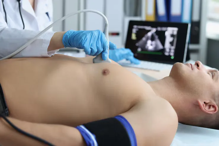
In a transesophageal echocardiogram (TEE), a slim ultrasound probe is passed through the mouth into the oesophagus (food pipe), which lies directly behind the heart. Because there are no ribs or lungs in the way, TEE gives exceptionally clear, close-up images of the atria, the atrial septum, the valves and the great vessels.
In children, TEE is performed under sedation or general anaesthesia so that it is comfortable and safe. It is most often used to guide catheter procedures such as ASD device closure, during heart surgery, and when a regular chest echo cannot answer an important question.
Superior Detail
Clearer views of the atrial septum, valves and small structures that are hard to see from the chest.
Procedure Guidance
Real-time imaging to measure defects and confirm correct device position during interventions.
Pediatric Probes
Small, child-sized probes allow TEE to be performed safely in infants and older children.
When Is It Recommended?
TEE is advised when detailed imaging will change the treatment plan, for example:
- Assessing the size and rims of an atrial septal defect before device closure
- Guiding ASD, PFO or other device closures in the cath lab
- Checking the result of heart surgery in the operating room
- Poor chest echo windows in older children, adolescents or after surgery
Not sure if your child needs this?
Share your child's reports and Dr. Bharath's team will guide you on the next step.
Conditions Covered
Heart conditions commonly assessed or treated with this service.
Atrial Septal Defects
Precise sizing of secundum ASD and its surrounding rims to decide on device versus surgery.
Sinus Venosus & Venous Anomalies
Defects near the venous connections that are often missed on chest echo.
Valve Disease
Detailed mitral, tricuspid and aortic valve anatomy before or after repair.
Infective Endocarditis
Detection of vegetations and abscesses on valves and devices.
Intracardiac Clots
Assessment of thrombus in the atria or around devices and conduits.
Post-Surgical Hearts
Evaluation of baffles, patches and residual leaks after congenital heart surgery.
How It Is Performed
What happens on the day, explained simply so you know what to expect.
- 1Step 01 / 05
Pre-Procedure Check
Review of fasting status, medical history and consent with the anaesthesia team.
- 2Step 02 / 05
Anaesthesia
Your child is given sedation or general anaesthesia so that the procedure is painless.
- 3Step 03 / 05
Probe Placement
A lubricated pediatric probe is gently guided through the mouth into the food pipe.
- 4Step 04 / 05
Imaging
Multiple views of the heart are recorded; during interventions, imaging continues throughout the procedure.
- 5Step 05 / 05
Wake-Up & Review
The probe is removed, your child recovers from anaesthesia and the findings are discussed with you.
Benefits, Risks & Care
A balanced overview, plus how to prepare and what recovery looks like.
Key Benefits
- Much clearer images than a chest echo in many situations
- Makes catheter device closures more precise and safer
- No radiation exposure
- Can avoid more invasive diagnostic tests
Every child is different. Specific risks, benefits and alternatives for your child are discussed in detail during consultation.
Why Families Choose Dr. Bharath A. P.
Super-speciality training, academic experience and hands-on cath lab expertise.
National Board super-speciality fellowship at Madras Medical Mission Hospital, Chennai.
Assistant Professor of Pediatric Cardiology at Sri Jayadeva Institute of Cardiovascular Sciences and Research since 2015.
Diagnostic catheterisation and interventions including ASD, VSD and PDA closures, valvuloplasties and stenting.
Co-author of peer-reviewed articles in Heart Lung Circulation, Archives of Disease in Childhood and Annals of Pediatric Cardiology.
Frequently Asked Questions
Answers to common questions about Transesophageal Echocardiography (TEE).
Why does my child need TEE if they already had an echo?
+
A regular echo gives most of the information needed, but some structures — such as the rims of an atrial septal defect — are seen much more clearly from the food pipe. TEE is advised only when this extra detail will guide treatment.
Will my child feel the probe?
+
No. Children are given sedation or general anaesthesia, so they are asleep and comfortable throughout the procedure.
Is TEE done separately or during the device closure?
+
It can be either. Sometimes TEE is done beforehand to plan treatment; more often it is performed during the catheter procedure under the same anaesthesia, so your child does not need a separate session.
When can my child eat after TEE?
+
Once your child is fully awake and swallowing normally, sips of water are started, followed by a normal diet — usually within a few hours.
Is TEE safe for small babies?
+
Yes, when performed with child-sized probes and under anaesthesia care. Breathing, oxygen levels and heart rate are monitored throughout, and the probe size is chosen to suit the child.
Will my child have a sore throat afterwards?
+
A mild sore throat is common for a day. Cool drinks and soft foods help, and it settles on its own without treatment.
Visit the Clinic
VEGA CURRE Multispeciality Clinic
2nd Floor, Shop No. 24 & 25, 3rd A Cross, Bhuvaneshwari Nagar, Dasarahalli Main Road, Hebbal Kempapura, Bengaluru – 560024