

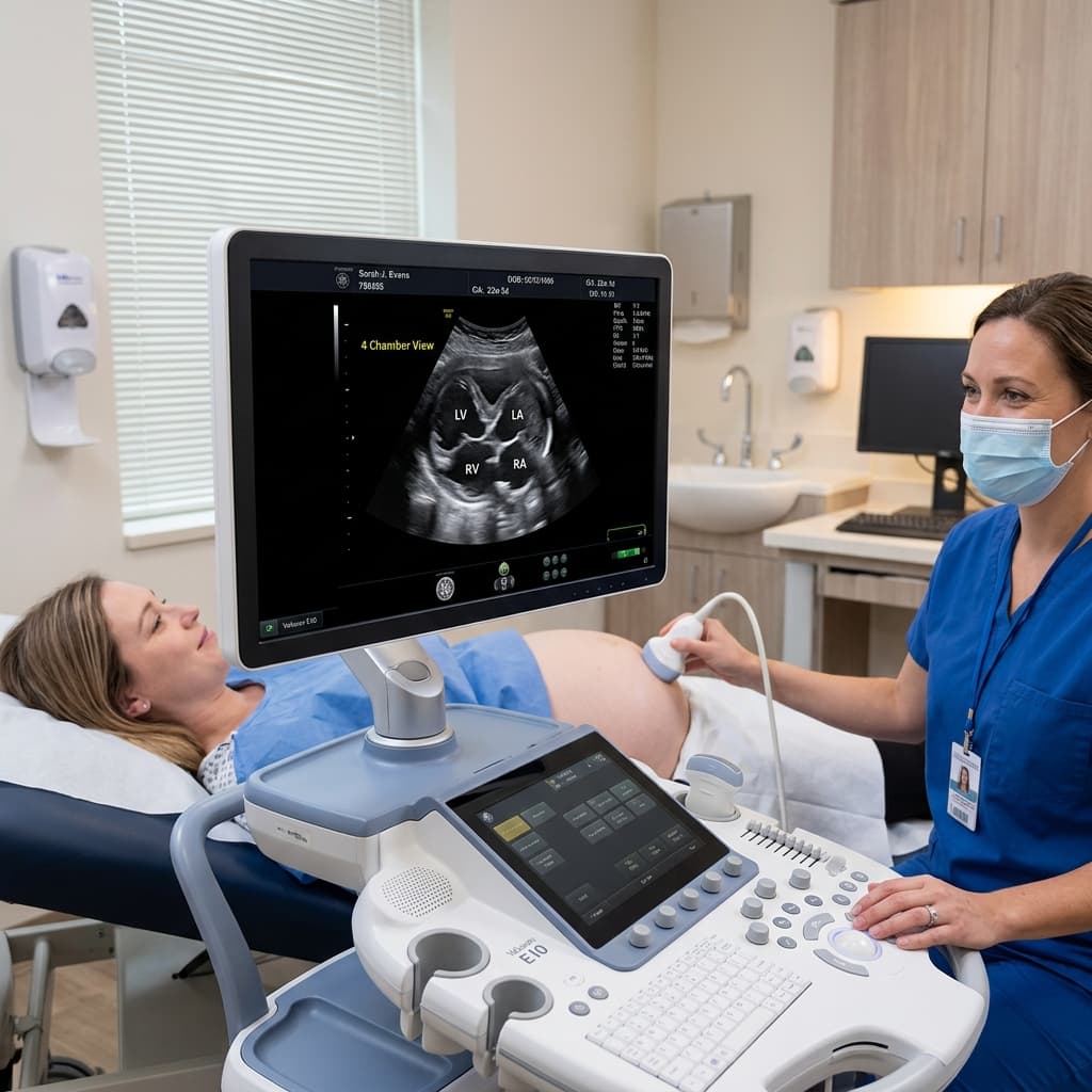
What is a Fetal Echocardiogram?
A fetal echocardiogram is a specialized ultrasound examination dedicated specifically to evaluating the developing heart of an unborn baby while still inside the mother's womb.
When is It Performed?
The optimal window for a detailed fetal echocardiogram is between 18 and 24 weeks of gestation. At this gestational age, the baby's heart structures and chambers are adequately developed to allow high-resolution visualization.
Common Reasons for Referral
- Sub-optimal or suspicious heart views on a routine 20-week target anomaly scan
- Maternal diabetes (pre-existing or gestational), autoimmune conditions like lupus
- Family history of congenital heart disease in siblings or parents
- Twin or multiple gestation pregnancies, or IVF conceptions
- Abnormal fetal heart rhythm (arrhythmias) noticed on routine checkups
Benefits of Prenatal Cardiac Diagnosis
Early identification of congenital structural heart conditions allows the medical team (pediatric cardiologist, obstetrician, neonatologist, and pediatric cardiac surgeons) to plan a safe, coordinated hospital delivery with immediate postnatal support in place.