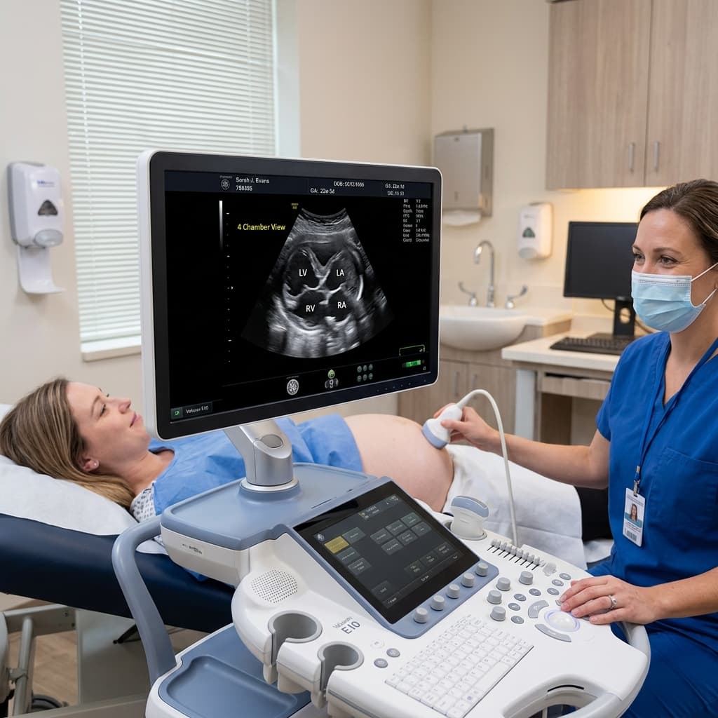


Understanding the Echocardiogram
A pediatric echocardiogram (often simply called an "echo") is one of the most important diagnostic tools in pediatric cardiology. It is essentially an ultrasound of the heart.
How Does It Work?
The test uses high-frequency sound waves to create moving pictures of your child's heart. It allows us to see the heart's structure, the valves, and how blood is flowing through the heart chambers and major blood vessels.
Is It Safe?
Yes. An echocardiogram is completely non-invasive, painless, and uses no radiation (unlike an X-ray or CT scan). It is very similar to the ultrasound a mother receives during pregnancy.
What to Expect During the Test
The procedure usually takes between 30 to 60 minutes. Your child will lie on an examination table, and the technician (or doctor) will apply a warm, water-based gel to their chest. A small probe, called a transducer, is then moved across the chest to capture images.
For infants and toddlers, keeping them calm and still is important for getting clear images. Parents are usually encouraged to stay right beside their child, perhaps offering a bottle, a pacifier, or watching a favorite video on a phone or tablet to help them relax.
Why Might Your Child Need One?
We typically perform an echo to investigate:
- A heart murmur detected by a pediatrician
- Unexplained chest pain or fainting
- A family history of congenital heart disease
- Concerns about heart rhythm or function
An echocardiogram provides immediate, detailed information, allowing us to quickly reassure parents if the heart is normal, or accurately diagnose and plan treatment if a condition is found.